[ Document Identification Number : DIN00032507 ]
Proceedings of the 2nd Symposium of the 'Color' of Digital Imaging in Biology and Medicine,
7.1-7.3, 2000.03.25
<http://biocolor.umin.ac.jp/sympo200004/proc17.pdf>
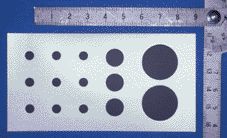
パネルディスカッション「デジタル生体医用画像の色のあり方」 ―その2:医療ニーズからのアプローチ― 形成外科領域における臨床写真のキャリブレーションの実際 寺田 伸一*1 (sterada@prs.twmu.ac.jp)、野崎 幹弘*1 *1東京女子医科大学形成外科 The 2nd Symposium of the 'Color' of Digital Imaging in Biology and Medicine Panel Discussion "What the 'Color' of Digital Imaging in Biology and Medicine Should Be" - Part 2 : Approaches From the Needs in Medical Practice - A Practical Method to Compensate Color Differences Among Pictures Taken for Case Records Shin-ichi TERADA*1 (sterada@prs.twmu.ac.jp), Mikihiro NOZAKI*1 *1Department of Plastic Surgery, Tokyo Women's Medical University Summary In plastic surgery, photographs have been intensively used for recording various lesions at the surface of the human body and the effects of plastic operations. Usually single-lens reflex cameras are used under fixed conditions to take standardized pictures, but this method is not properly used for digitized images taken by digital cameras which are of late rapidly coming into use A color management system for these digitized photographs has been devised and put into practice by us. In this system, a round shaped gray seal (G-ROUND18, Kyowa Tokei Kogyo, Fig. 1) which has a color value of '49.5, 0, 0' represented by the L*a*b* system is used as a color calibrator. To evaluate the system, (1) 10 pictures of this calibrator were taken on reversal film by a single-lens reflex camera (EOS650, 50mm Macro lens, Macro ring lite, Canon) by stopping the lens intentionally, (2) these pictures were digitized by a film scanner (Picture MD Writer, Konica) and color compensation made by using the plug-in Photoshop(TM) software (Gray Balance, Konica), (3) color compensation was made using a built-in filter of the film scanner on the same image files, and (4) color values represented by the L*a*b* system of this calibrator and image files made at (1), (2) and (3) were measured using a spectrophotometer (CR-221, Minolta). The results were '47.5+-0.07, -1.11+-0.04, 0.1+-0.02' (N=10), '26.6+-0.7, 9.0+-2.1, -14.8+-2.4', '25.1+-0.9, -1.1+-2.8, 0.6+-1.6' and '47.2+-0.8, 0.7+-1.8, -0.8+-2.5'. This results suggests that our method for color compensation is very effective in precise digitization of photographs with a film scanner. |
はじめに 形成外科は従来より熱傷や指切断などの外傷からあざや唇裂口蓋裂、小耳症、漏斗胸などの先天異常、悪性腫瘍切除後の再建手術など多岐にわたる体表疾患を治療対象としている。このため、医学ドキュメントとして臨床写真を頻繁に撮影している。一般的には一眼レフカメラに50mmマクロレンズとリングフラッシュを装着して、1:3、1:5、1:7,1:9などの固定視野で撮影サイズを一定にして、対象別の規格撮影を行ってきたが、近年の高画質デジタルカメラとパーソナルコンピュータの急速な普及から、フィルム現像が不要となるデジタルカメラへの移行が始まっている。しかし、安価なデジタルカメラでは一眼レフ機能が削られているため、画像サイズの不一致や色調のばらつきが目立つようである。そこで、われわれは一定サイズの18%反射率のグレースケールを基準としたカラーマネージメントシステムを開発し、臨床写真のキャリブレーションに利用しているので、報告する。 方法 キャリブレーションとして被写体と共に18%反射率を有するグレースケールの直径5mmの円形シール(G-ROUND18、協和時計工業、Fig. 1)を貼付した。このシールのL*a*b*の理論値は49.5、0、0である。これを色彩色差計(CR-221、ミノルタ)で計測した。次にグレースケールを配置したモデルを一眼レフカメラ(EOS650、50mmMacro lens、Macro ring lite、キヤノン)でリバーサルフィルムに撮影した。この際、レンズの絞りを-2から+2に変化させた。フィルムを現像した後、フィルム連続スキャナ(ピクチャーMDライター、コニカ)でデジタル化した。Adobe Photoshop上でLab画像に変換し、グレースケールのL*a*b*を計測した。色調補正はグレースケールに基づき、スキャナ内蔵の補正フィルタおよびPhotoshop上の補正プラグイン(gray balance、コニカ)を用いた。 |
|
結果 色彩色差計によるグレースケールのL*a*b*値は各々47.5±0.07、-1.11±0.04、0.1±0.02 (N=10)であった。リバーサルフィルム(Konicachrome)で‐2絞りで撮影した画像は補正不能であった。‐1絞りでは元画像が26.6±0.7、9.0±2.1、-14.8±2.4であった。Photoshop上で補正した画像では25.1±0.9、-1.1±2.8、0.6±1.6であった。一方、スキャナ上で補正した場合は47.2±0.8、0.7±1.8、-0.8±2.5であった。 結語 スライド写真でもスキャナで読み込む際には色調補正を行った方が色再現性の点で、良好な画像が得られた。グレースケールによるカラーマネージメントシステムの有用性が示唆された。 |