[ Document Identification Number : DIN00032506 ]
Proceedings of the 2nd Symposium of the 'Color' of Digital Imaging in Biology and Medicine,
6.1-6.3, 2000.03.25
<http://biocolor.umin.ac.jp/sympo200004/proc16.pdf>
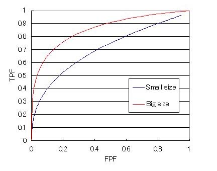
Summary Images of an artificial lesion made on human skin were captured and digitized by a digital camera at two kinds of spatial densities of pixels and the differences of their diagnostic reliability were examined with the ROC (Receiver operating characteristic) analysis. Hand skins of 13 humans were marked by red ink with a fiber-tipped pen of 0.4mm diameter and 50 pictures were taken with CAMEDIA-XL (C-1400, Olympus Co., Ltd.) in a predetermined condition before and after marking. These images were digitized into two kinds of format, 1280x1024 pixels (large format) and 640x512 pixels (small format), then compressed into the average ratio of 48.9% (the maximum ratio being 38.8% and the minimum 63.3%.) These image files were displayed under a determined condition and observed by eight staff members of Department of Medical Informatics of Hokkaido University (male: 5, female: 3) twice with at least a one-week interval. The computer software used for ROC analysis is ROCKIT (Chicago University.) The result is shown in Fig. 1. The As values are 0.70+-0.06 for the small size and 0.86+-0.03 for the large size. This result suggests that the artificial lesion recorded as a digital image will be diagnosed differently according to the spatial densities of pixels composing the image. |
1.目的 市販のデジタルカメラを臨床に活用することは、表在性病変の所見の伝達と診断に有効であると考えられる。今までに、デジタルカメラの活用例に関する報告は多くあるが、デジタルカメラで得られた画像の画質、読影と判断に関する定量的な研究はあまりなされて来なかった。今回、我々はデジタルカメラで撮影された色画像の画像サイズが読影判断に及ぼす影響を検討するため、皮膚上の赤色点を疑似病変と見立て、疑似病変の識別と判断に関してROC(Receiver operating characteristic)曲線による基礎的な実験を行った。実験方法など検討の余地があるが、その実験方法と得られた結果について報告する。 2.方法 2.1 実験装置・実験試料 撮影環境を一定にするために照明用白色蛍光灯80Wを2本を備え、壁内面を白色の壁紙を貼った70×70×80cmの撮影用箱を作成し、撮影は全てその撮影箱の中で行った。デジタルカメラは、オリンパス製を使用した。撮影条件は、画質モードをSHQ、ホワイトバランスをAUTOとした。フラッシュは使用せず、露光はスポット測光モード(自動)、オートフォーカスとした。撮影距離は80cmとした。 実験試料は、手関節より先の手部のデジタル画像とし、無信号画像(手部を普通に撮影したもの)50枚と有信号画像 (中手指節関節と手関節の間に疑似病変として0.4mmの赤色水性ペンにより点をつけたもの) 50枚を用意した。手部の画像はボランティア13名(男性7名、女性6名、平均年齢±標準偏差:27.7±5.7歳)の手部を用いた。全ての画像について、サイズ大(1280×1024)及びサイズ小(640×512)の2種類のサイズの画像を作成した。これらはJPEG圧縮されており、平均圧縮率は48.9%(最小38.8%、最大63.3%)であった。 2.2 読影実験 医療情報部員8名(男性5名、女性3名)を読影者とし、連続確信度法によるROC曲線により評価を行った。読影環境は太陽光による影響を防ぐため、窓がなく蛍光灯のみの部屋を使用した。読影には、NEC社製CRTディスプレイ(DV17B2)を用い、ディスプレイの設定は32bit、1024×768とした。読影時の照度はディスプレイ前40cmの位置で360lxとなるようにした。画像表示ソフトウェアはPhotoDog(フリーソフト)を、ROC解析にはROCKIT(シカゴ大学製)を使用した。読影は2回に分けて行い、少なくとも1週間以上間隔を空けるようにした。 3.結果 得られた結果をFig. 1に示す。ROC曲線から得られたのAz値(平均値±標準偏差)はサイズ小で0.70±0.06、サイズ大で0.86±0.03であった。2群間のpaired-t検定の結果はp<0.01となり2つ画像グループ間に有意差が認められた。この結果より微小(0.4mm)の赤色疑似病変の読影と判断には、画像サイズが影響していると考えられる。画像サイズは画像保管や画像伝送に大きく影響するが、病変の大きさと種類により適切な画像サイズを選択する必要があろう。今後、本実験も問題点(疑似病変の性状や評価者の特性)を検証し、更に画質を変化させた実験を行いたいと考えている。 |
|