[ Document Identification Number : DIN01022802 ]
Digital Color Imaging in Biomedicine (in press), 2001.02.28 (draft)
<http://biocolor.umin.ac.jp/book200102/din01022802.pdf>
Digital Color Imaging in Biomedicine (in press), 2001.02.28 (draft)
<http://biocolor.umin.ac.jp/book200102/din01022802.pdf>
- A noncontact-type spectrocolorimeter with a globe for light integration - Akiko UCHIDA*1 (auchida@md.tsukuba.ac.jp) *1Department of Plastic Surgery, Institute of Clinical Medicine, University of Tsukuba |
Abstract: Standard instruments used for measuring skin color have some problems: 1) they are affected by pores and wrinkles in the skin, 2) they are affected by various illuminant conditions, 3) the edge loss error caused by the semitransparency of skin. We have developed a new measuring system for the color of skin based on a non contact type color spectrometer equipped with a globe for light integration, and succeeded to improve these problems remarkably. |
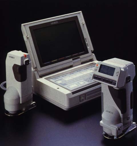
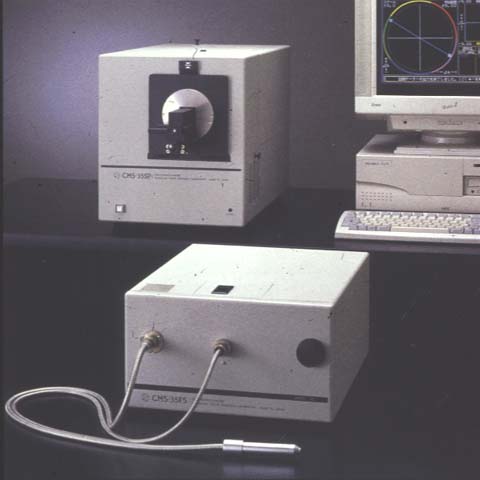
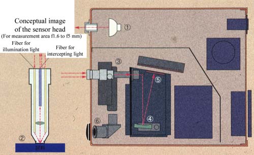
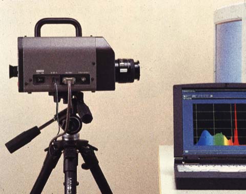
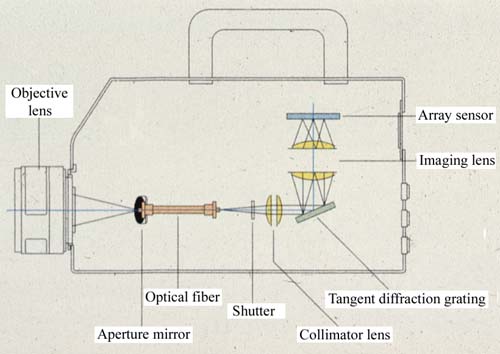
1. Introduction Conventionally, skin color is often measured with a contact type spectrophotometer usually used for measuring the color of a physical object (Fig. 1-6). Although it contains an illuminating unit and ensures high reproducibility, the contact type spectrophotometer is not suitable for measuring the color of skin which has an indented surface, hairs, or a sore with wet secretion, or the color of an angioma which varies in color depending on the pressure applied to it. On the other hand, the non-contact type color spectrometer (Fig. 7,8) usually used for measuring the color of a light source cannot produce a constant illumination, nor offer sufficient precision to comply with international standards on the measurement of the color of a surface. We therefore developed a non-contact type color spectrometer for measuring the color of skin, the illumination and precision of which are similar to those of the contact type. This paper introduces the non-contact type color spectrometer that we manufactured. |
|
|
| |
|
|
|
| |
|
|
|
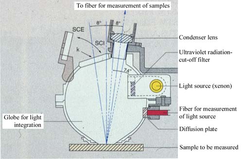
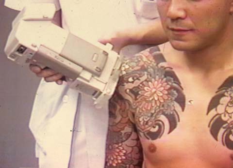
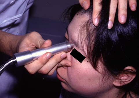
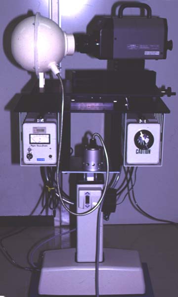
2. Methods An 8-inch globe for light integration is attached to the aperture of a spectroradiointensimeter (CS-1000, Minolta). A 150-watt xenon fiber light source was inserted. The tip of the globe for light integration is opened approximately 5 cm to the target, or skin surface. The center of the measured area (4 mm diameter) is not in any contact such as with the transparent plates (Fig. 9). Using this device, skin of various conditions was measured and reproducibility was evaluated. Measured values were compared with those obtained by contact-type colorimeters (CM-2022, Minolta; CMS-35FS, Murakami Color Technology). |
| |
|
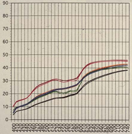
3. Results (Fig. 10) Compared with the two contact-type instruments, reflectance curves of the noncontact-type instrument with a globe were sharper. The reproducibility of data in repeated measurements was highest in the order of contact-type CMS-35FS > noncontact-type instrument with a globe > contact-type CM-2022. When flared-up skin several days after sunburn was measured, only the noncontact-type instrument with a globe measured significantly high reflectance, particularly that of the long wavelength area. |
| |
 (b) 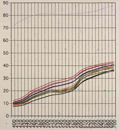 (c) 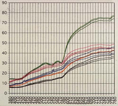 |
4. Discussion When semitransparent skin is measured by a conventional contact-type colorimeter, because of the partial lack of reflected light inside the skin, measured values tended to have reduced luminosity and saturation at peripheral portions of measured areas (edge loss error). Because the noncontact-type instrument with a globe has a sufficient illumination area (diameter 50 mm) relative to the measured area (diameter 4 mm), the edge loss error can be eliminated. Consequently, skin color that is more natural and closer to that visually observed is reproduced. Furthermore, the globe for light integration products a perfectly radiant distribution of illumination. Because reflected light is measured, conditions are closer to those of visual observation than those of 45-degree illumination or ring illumination, and stability is also higher. The CMS-35FS is a pen-type hand piece. It is easy to hold the instrument steadily and to measure areas. The measuring time is only 3 seconds. With the other two instruments, minor tremors of instruments and patients produced a shift in measured areas. Measurement of skin color several days after sunburn was affected by optical factors in the cortical layer such as angiotelectasia of the dermis, appearance of sunburn cells in the epidermis, increased secretion of melanin granules, activation of division of epidermal cells and ablation of the cuticle. It is inferred that the reason why values of sunburned skin measured by the noncontact-type instrument were markedly different from those measured by other types of colorimeter is that the former instrument has a structure that can intercept spectral light in the skin cortex as mentioned above. We plan to conduct experiments on depth and directional disassimilation. 5. Conclusion This measuring system improved the dispersion of measured values caused by skin conditions such as wrinkles and pores, which are a problem with conventional contact-type colorimeters. Noncontact-type colorimeters used to have the weakness of being easily influenced by the measurement environment. The attachment of a globe for light integration has solved this problem by producing a radiant distribution of illumination and light sources with changeable intensity. The new instrument eliminates the problem of fading of skin capillary due to pressure on the measured area, as well as the problem of edge loss error in the measurement of color of semitransparent objects. We plan to conduct further studies and obtain reliable data on the size of the globe for light integration, intercepting angle, intensity of radiation light and improvement of data reproducibility by repeated measurements to achieve optical conditions that meet JIS. |